Pages

Tuesday, April 15, 2014

Jack Frost Does Not Work Alone


Dead canes of a flower carpet rose
As March was going out like a lamb, a nursery submitted four container-grown shrubs to the PDIC: three rose cultivars and a lilac. Very young shoots on these plants were withering and dying. At least in the case of the lilac – and possibly with the roses, too – the new flush of growth had been hit by the last freezes of the spring. While you’d expect the tender shoots to be blasted by the cold, in this case the woody stems were also dying. Bacterial streaming was seen in much of the stem tissue. We don’t see fire blight on rose or lilac, so what was happening?

The grower suspected Pseudomonas blight. He was right.

A bacterium and its victims

Bacteria were cultured from the stem tissue of the affected plants. Since only Pseudomonas species were of interest, only colonies fluorescent* on a special agar medium were chosen for further work-up. Unfortunately there are a lot of nonpathogenic (non-disease-causing) Pseudomonas species in this world, so it took a little time to sift through the isolates and confirm the diagnosis as Pseudomonas syringae.

Wilting new shoot of a container-grown lilac.
Although Pseudomonas syringae is named after lilac (Syringa), it is capable of causing cankers and dieback in a wide variety of plants. Besides lilac, we’ve found it on the following woody ornamentals: cherry-laurel, flowering quince, Indian hawthorn, Yoshino cherry, and multiple varieties of rose.  In addition, we’ve recovered it from leaf spots of hydrangea and Japanese holly. Bacterial canker caused by Ps. syringae can be a serious problem in peach orchards, but with woody ornamentals we almost always see it in nursery situations. One exception came in last year, on the twig of a weeping willow from a home landscape. As the weather warms up and cankers become inactive, this disease becomes more difficult to detect. According to the PDIC's records, almost every case of Pseudomonas bacterial canker on woody ornamentals since 2008 was diagnosed between February and May. The bacterium is still present on and within plants during the summer, but what I believe is happening is that the hotter temperatures slow down this particular bacterium at the same time as they're (up to a point) invigorating most plants, thus shutting down the disease process temporarily.

Note: We occasionally find Ps. syringae causing leaf spots on ornamentals in the greenhouse, and there are variants – called pathovars – that cause certain very specific problems such as bacterial speck of tomato and angular leaf spot of cucurbits.

How Pseudomonas syringae does its dirty work

Blighted shoots and a Pseudomonas stem canker on rose
Like many bacteria, Pseudomonas syringae is able to live and multiply on plant surfaces. This is known as its epiphytic (“on the plant”) phase. In the recent case, the bacteria were almost surely present before the spring flush occurred, and so were able to strike quickly. These bacteria enter plants following injury, in particular frost damage. What’s more, the bacterial cells actually promote freeze damage through a process known as ice nucleation. How this works is succinctly expressed by Sinclair and Lyon:

"Ice-nucleating strains of P. syringae and certain other bacteria can trigger ice formation in plant tissues cooled to between -2 and -5ºC [28 to 23 ºF] but not acclimated to low temperature. Ice then disrupts cells, causing symptoms of frost damage. In the absence of an ice-nucleating factor, frost-sensitive plants may tolerate brief cooling to these temperatures because water in their tissues remains in liquid form, supercooled." (Diseases of Trees and Shrubs, 2nd Ed. 2005. p.368)

For more information, see this review by Gurian-Sherman and Lindow. You might also check out this laboratory video of ice nucleation by bacteria added to supercooled water.

As if this ice-nucleation trick were not enough, Pseudomonas syringae also produces a toxin that damages plant cells.

How to reduce your losses

The most important way to minimize damage to woody plants from Pseudomonas syringae is to limit the stressors that predispose plants to infection. Stress factors include pruning injury and frost injury. Bacterial canker of stone fruits caused by Pseudomonas syringae can be reduced by pruning in the early summer, instead of the fall or winter. Sanitize shears or knives frequently, and avoid working the plants when wet. Don't overfertilize plants, especially when they need to harden off for the winter. Protect plants during cold snaps. Don't allow plants to undergo stress from too much or too little water. Keep foliage and stems as dry as possible by changing irrigation methods or reducing overhead irrigation, which favors and spreads the bacteria. If you’ve already had this problem, Ps. syringae is probably present as epiphytic populations on the surfaces of much of your nursery stock and even the surrounding weeds. There are few chemical options that hold any promise, at least not enough to make a recommendation.

As I write this, winter is getting ready to take one last shot at North Carolina, with freeze warnings up for the western half of the state. It's another opportunity for Pseudomonas syringae, too.

*Chemist’s Corner:

Colonies of fluorescent pseudomonads photographed under UV light.
The fluorescent pigments of Pseudomonas species are seen by shining a long-wave UV lamp on the cultures. These compounds belong to a class of chemicals called siderophores. If you know Latin, you might think that siderophore means “star bearer”, but in this case the root is the Greek word for “iron”. (A big thanks to Roland Wilbur Brown’s 1956 book Composition of Scientific Words for setting me straight.) Iron is an essential element for microbial growth, and siderophores have the important task of scrounging precious iron from the bacteria’s environment.

Thursday, April 3, 2014

A. destructor

This may look like the toothy mouthparts of some sinister little animal,
but it's really the posterior end of an armored scale (Diaspididae).
UPDATE: After looking at new scales on an Aucuba recently (July 5 2016) it was determined that the scales identified in this post as Aspidiotus destructor, are most likely Aspidiotus hederae, the oleander scale. All information on the former species and on armored scales remains correct, but note that the images of the scales represent the latter species.

Armored scales (Diaspididae) are one of the most common insect pests of ornamentals. More like diseases than insects, these sedentary bugs (literally - they are in the Order Hemiptera) sit and suck the sweet fluids of plants, all the while taking energy from their host and replicating as big sacs of eggs. Their babies, called "crawlers", infest new areas and settle in for the long haul of motherhood (or the short, but free, life of a winged male).

Over the past month or so we received two samples of different plants that had an interesting armored scale infestation. The first was poet's laurel (Danae racemosa), that looked as if it was a variegated variety from the amount of chlorosis associated with the scales:

Poet's laurel (Danae racemosa) leaves with yellow and brown areas due to scale insect pressure. 

Looking under the microscope, I noticed two types of armored scales. The first, and less common, were some typical brown, oyster-shaped fern scales (Pinnaspis aspidistrae). However, the most noticeable scale was one I did not recognize. It had a very thin, translucent test that resembled delicate wax paper with a bright yellow scale underneath:

Although it may look like these scales are under the epidermis of the plant, they are really hiding under a thin test (the covering common to armored scales, to which they owe their name).

When lifted off, the scales underneath looked like this:

Scales with their test removed (top one is facing right and bottom one is facing left).

The scale certainly had the rounded shape characteristic of most members of the subfamily Aspidiotinae (as opposed to the elongate shape of most Diaspidinae, such as the previously mentioned fern scale)...but what was it? Well, I have to confess that our friend and former clinic member Dave Stephan took a peek during a visit and thought it looked like the genus Aspidiotus. Luckily he said that, because the scale book I most often reference first, Scale Insects of Northeastern North America by Michael Kosztarab, does not cover this genus which is primarily Southern in distribution. So I referenced another great resource Ferris's Atlas of the Scale Insects of North America. Lo and behold, Dave was right - the scale keyed out to the genus Aspidiotus and further to the species A. destructor, the coconut scale.

Luckily I had identified that sample, because the same day there was an image of aucuba (Aucuba japonica) that was submitted with a potential scale infestation. Although I was not able to ID the species from the pictures, when the sample came in I recognized the similarities with A. destructor:

The yellow spots on this aucuba leaf are intentional variegation - not chlorosis attributed to the scales. 
Close up of different-sized coconut scales (Aspidiotus destructor) oleander scales (Aspidiotus hederae).

After clearing some specimens I was able to definitively ID them as the same scale. Was this a coincidence? Probably, but who knows whether these scales are becoming more abundant. Our clinic records show that A. destructor was only submitted and identified four times in the 14 years prior to these two samples. Does that mean that we will be seeing more of this scale? I am not ready to conclude that, but if more are submitted this year we may have to investigate what's going on.

----------

A little more on the scale.

Aspidiotus destructor was described by Signoret in 1869 and goes by several common names including bourbon scale and transparent scale (I am assuming based on the thin test). The scale appears to be Southeast Asian/Pacific in origin, but has been spread throughout the world. Although mainly a pest of coconut and banana, it is extremely polyphagous being found on over 60 families of plants. The genus is characterized by the following traits that can be seen in the title image (Ferris, 1938):

  • absence of paraphyses or intersegmental scleroses
  • three pairs of lobes with no indication of a fourth pair
  • plates long, flat and fringed (two between median lobes, two between median and second lobes, three between second and third lobes, and a variable number beyond third lobe)
  • characteristic sclerotization on dorsum of pygidium

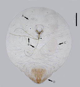
The scale can cause significant economic damage at high densities (which can be common), stunting plants and eventually killing them if enough of the leaves become unable to undergo photosynthesis. Treatments can include chemical control, but there is also a diversity of natural enemies known to attack the species including various fungi, ladybugs, thrips, mites and several parasitoid wasps, most of which are in the family Aphelinidae. In fact, after clearing the scales from the aucuba, I noticed some sinister-looking aliens inside a few of the scales that are certainly wasp larvae and likely a species of Aphelinidae (below "A"):


A cleared female coconut  oleander scale showing a parasitoid wasp larva (A), mouthparts/stylets (B), scale egg (C)
and pygidium (D). [Thanks to Mike Munster for helping take pictures of the scale under the microscope]

I don't know whether or not the wasps are able to keep these scales in check alone, but there were at least a few  being eaten by these tiny larvae. Every bit helps I guess!

References:
Ferris, G.F. 1938. Atlas of the scale insects of North America. Series 2. Stanford University Press, Palo Alto, California

Signoret, V. 1869. [Essay on the gall forming insects (Homoptera - Coccidae) - 3rd Part.] Essai sur les cochenilles ou gallinsectes (Homoptères - Coccides), 3e partie. Annales de la Societe entomologique de France (serie 4) 9: 97-104.